Frown lines and the glabellar complex
Anatomy of corrugator and procerus, the mechanism of eyelid ptosis after glabellar treatment, and why this region leads the reported blindness series.
The glabella is the region between the eyebrows, above the root of the nose. Vertical and transverse lines here are produced by repeated contraction of a small muscular complex, and the area is treated more often than almost any other in aesthetic practice. It is also the most dangerous single region on the face for injected material, and those two facts sit uncomfortably together.
Relevant anatomy
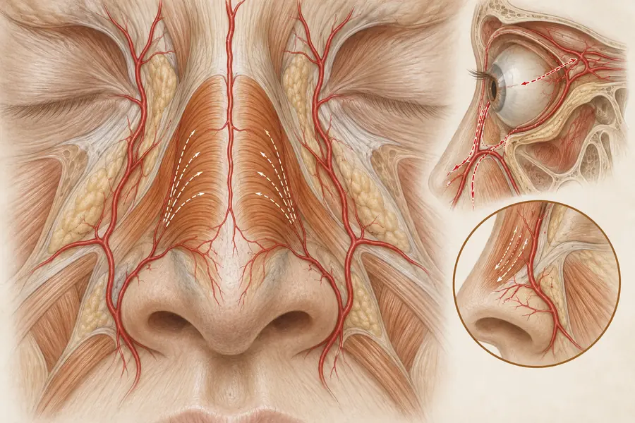
The corrugator supercilii arises from the medial supraorbital rim and passes superolaterally to insert into the dermis of the mid-brow, drawing the brows medially and downward and producing the vertical lines. The procerus runs from the nasal bones and upper lateral cartilage to the skin of the lower forehead, pulling the medial brow down and creating the transverse line across the root of the nose. Depressor supercilii and the medial fibres of orbicularis oculi contribute further depression, and frontalis opposes all of them.
The supratrochlear and supraorbital arteries emerge from the orbit at the medial and mid supraorbital rim respectively. Both are terminal branches of the ophthalmic artery, itself the first branch of the internal carotid. This is the critical point: the glabellar vessels are in direct continuity with the retinal circulation, and they are small, superficial and close to the periosteum in a region with limited soft tissue depth.
Mechanism of eyelid ptosis
Levator palpebrae superioris lies within the orbit and elevates the upper eyelid. It is not a target of glabellar treatment, but toxin placed low or in excessive volume near the medial supraorbital rim can track through the orbital septum and weaken it, producing true eyelid ptosis. This is distinct from brow descent, which follows frontalis weakening and presents as a heavy or lowered brow with a normal lid margin. The distinction matters because the two have different courses and different implications for how an unhappy outcome is interpreted.
Documented risks in this region
Reviews of filler-induced blindness consistently place the glabella at or near the top of the reported case series, with the nasal region close behind. The mechanism is retrograde embolisation: material forced into a terminal branch at pressure exceeding systolic travels backwards along the supratrochlear or supraorbital artery towards the ophthalmic artery, then passes anterograde into the central retinal or posterior ciliary vessels when injection pressure falls. Visual loss is typically immediate and painful, frequently accompanied by skin changes in the same vascular territory, and published outcomes for recovery of vision are poor. Guidance on the management of hyaluronic acid vascular occlusion treats this as a time-critical emergency and assumes reversal agent is immediately available wherever hyaluronic acid is used.
Where assessment goes wrong
The recurrent errors here are anatomical and procedural rather than aesthetic: treating the glabella as a routine, low-consequence area precisely because it is treated so often; failing to account for previous surgery, scarring or prior filler altering vessel course and tissue planes; assuming that surface landmarks reliably predict arterial position when cadaveric studies show substantial variation between individuals and between sides of the same face; and failing to separate brow descent from true ptosis when reviewing a poor result, which leads to the wrong conclusion about what happened.