Smokers lines and perioral rhytids
Orbicularis oris as a sphincter, the labial arteries near the wet-dry border, and the functional cost of weakening a muscle used for speech and continence.
Perioral rhytids — the vertical lines radiating from the vermilion border, colloquially and inaccurately called smokers’ lines — arise from the interaction of repeated sphincteric contraction, dermal thinning, photodamage and loss of underlying support. Smoking accelerates them but is not required to produce them; they occur in lifelong non-smokers and are strongly associated with sun exposure and with skin phototype.
The muscles around the lip
Orbicularis oris is the sphincter itself, encircling the mouth. Into it insert the muscles that move the lip, and one of them answers a question that comes up constantly: the muscle that surrounds the upper lip, elevates it and dilates the nostrils is the levator labii superioris alaeque nasi. Its name describes the action exactly: elevating the lip of the upper jaw while dilating the wing of the nose — the longest muscle name in the body, and the reason a gummy smile and a flaring nostril are related problems.
Relevant anatomy
Orbicularis oris is not a simple ring. It comprises deep fibres derived from buccinator that run circumferentially and are responsible for the tight seal against the teeth, and superficial fibres inserting into the dermis and vermilion, responsible for pursing and eversion. Peripheral fibres interdigitate at the modiolus with zygomaticus major, risorius, levator anguli oris, depressor anguli oris and buccinator, so the muscle is functionally continuous with the entire perioral group rather than isolated from it. The dermal insertions of the superficial fibres are why sustained contraction produces radial lines running perpendicular to the vermilion.
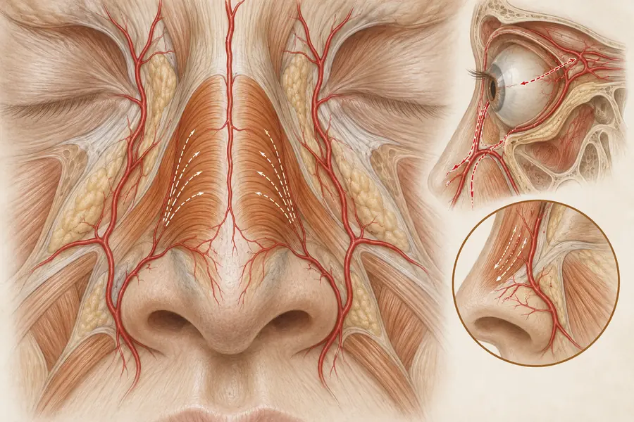
The superior and inferior labial arteries branch from the facial artery near the oral commissure and run circumferentially within or deep to the muscle, typically near the wet-dry border rather than at the vermilion edge, though cadaveric studies report considerable variation in both depth and position between individuals. Published danger zone mapping and safe zone guidance both single out the perioral region for this unpredictability. Risk factor analyses of vascular occlusion record the lips among the more frequently implicated sites, in part because of anatomical variability and in part because the region is treated so often.
What the approaches aim to do
Two distinct rationales apply. Volumising approaches restore the vermilion border, the philtral columns and the structural support beneath the cutaneous lip, on the basis that lines form where dermal support has been lost. Relaxation approaches aim at reducing the amplitude of superficial orbicularis contraction so that the dermal insertions pull less forcefully. Resurfacing and energy-based approaches address the dermal component itself. The lines are usually a composite, and a plan that addresses only one contributor produces partial change.
Functional cost of weakening the sphincter
Orbicularis oris is a working muscle with daily functional obligations, and this distinguishes the perioral region from the upper face where the muscles treated are essentially expressive. Weakening it can impair the labial consonants of speech, drinking through a straw, whistling, playing a wind or brass instrument, and oral competence with liquids. These are not rare complications but predictable consequences of reducing sphincter strength, and they scale with how much function is removed. Anyone whose occupation depends on precise lip control is treating a working tool.
Where assessment goes wrong
Errors are conspicuous here in a way they are not elsewhere, because the lips are the region observers watch during conversation. Assessment fails when static lines are treated as though they were dynamic, so relaxation is applied to lines that persist at rest and remain visible with function additionally impaired. It fails when the vermilion border is overfilled and the philtral architecture flattened, producing the shelf-like appearance that is immediately identified by lay observers. It fails when asymmetry of the vermilion or the philtral columns is not documented beforehand. And it fails when the region’s vascular variability is assumed away on the basis of previous uneventful treatments.