Bunny lines on the nose and the perinasal danger map
Nasalis and bunny lines as compensation after glabellar treatment, and why the nasal region ranks second in the reported filler blindness series.
Two quite separate subjects meet at the nose. One is a minor dynamic line pattern of the nasal sidewall; the other is the second most dangerous territory on the face for injected filler. They are discussed together only because they share an anatomical region, and confusing the risk profile of one for the other is itself an error.
Bunny lines and nasalis
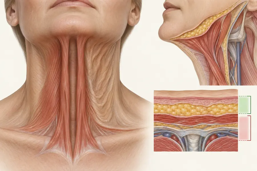
Bunny lines are the oblique rhytids that appear across the nasal dorsum and sidewalls on wrinkling the nose. They are produced chiefly by the transverse part of nasalis, which arises from the maxilla lateral to the nose and passes across the dorsum as an aponeurosis continuous with its fellow, compressing the nasal aperture. The procerus and the levator labii superioris alaeque nasi contribute at the root and the alar base respectively.
Their most clinically interesting feature is that they frequently appear or intensify after glabellar treatment. When corrugator and procerus activity is reduced, the expressive effort formerly delivered by those muscles is partly redistributed to adjacent unaffected muscles, and nasalis is the nearest available one. A patient who did not have bunny lines before glabellar treatment and has them afterwards has not developed a new problem; they are demonstrating recruitment. Anticipating this at the point of initial assessment, and explaining it, avoids the more common sequence in which the patient interprets the change as a treatment failure.
Why the nasal region is dangerous
Published reviews of filler-induced blindness spanning a century of case reports place the nasal region second only to the glabella in reported frequency. The vascular explanation is the anastomotic richness of the area. The dorsal nasal artery is a terminal branch of the ophthalmic artery and reaches the nasal dorsum after emerging above the medial canthus. The lateral nasal and angular arteries arise from the facial artery, part of the external carotid system, and anastomose freely with the dorsal nasal and with the columellar supply. This means external carotid territory communicates directly with internal carotid territory at the nose, and material entering a branch under pressure has a route to the retinal circulation.
The nose compounds this. Its skin envelope is tight and unyielding, particularly over the dorsum and tip, so injected volume raises local tissue pressure quickly and the margin before compromise is narrow. It is a common site of previous surgery, and prior rhinoplasty distorts both vascular course and tissue planes unpredictably. Danger zone literature accordingly lists it among the highest-risk sites, while more recent anatomical work has attempted to redefine the perinasal region in terms of safer corridors rather than blanket avoidance, mapping where the major vessels are least likely to run.
Documented risks
Reported events include visual loss, which is typically immediate; skin necrosis of the alar rim, dorsum or tip, following the territory of the occluded vessel; and, in the pooled case literature, ophthalmoplegia and cerebral infarction. Alar necrosis is the classic nasal presentation and can follow either direct arterial occlusion or compression from volume in a confined space.
Where assessment goes wrong
The errors are consistent: treating nasal filler as a minor procedure because the quantity involved is small; failing to elicit a history of previous rhinoplasty or filler; assuming surface landmarks predict vessel position in a region where anastomotic variation is the rule; and treating the low-consequence bunny line question and the high-consequence structural question as if they belonged to the same category of decision.