Platysmal bands and turkey neck
Platysma anatomy, what relaxation can and cannot achieve, why skin laxity is not a toxin problem, and the risks of deep or excessive neck treatment.
Vertical cords running down the anterior neck, prominent on speaking, grimacing or straining, are the free medial borders of the platysma. The colloquial description of a “turkey neck” conflates several different findings — banding, skin laxity, submental fat and loss of the cervicomental angle — and the first task in assessment is to separate them, because only one of them is muscular.
Relevant anatomy
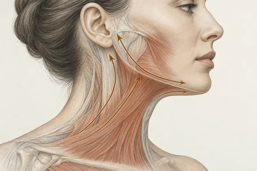
The platysma is a broad sheet within the superficial cervical fascia, arising over pectoralis major and deltoid, ascending obliquely across the clavicle and neck, and inserting into the inferior border of the mandible, the modiolus, the lower lip depressors and the skin of the lower face. It is innervated by the cervical branch of the facial nerve.
Its midline behaviour varies. Anatomical descriptions recognise several decussation patterns: fibres that interdigitate widely across the midline below the chin, fibres that meet only near the symphysis, and fibres that do not decussate at all. Where decussation is limited, the medial borders are free, and with age and loss of overlying fat these free borders stand forward as visible cords. This is why banding is prominent in some individuals and absent in others with comparable ageing.
Sensation over the anterolateral neck and the angle of the mandible comes from the cervical plexus, chiefly the transverse cervical and great auricular nerves, which emerge around the posterior border of sternocleidomastoid at the nerve point. These are sensory only; the motor supply to the platysma is facial, not cervical, and confusing the two leads to incorrect reasoning about what a given complication implies.
What relaxation can and cannot achieve
Relaxing the platysma reduces the tone in the band so that the cord no longer stands proud, and reduces the downward pull the muscle exerts on the jawline. Within its own terms this works, and the effect on visible cording during animation can be marked.
What it cannot do is tighten skin. Dermal laxity is a consequence of collagen and elastin loss, photodamage and volume change beneath. A relaxed muscle beneath lax skin may leave the skin looking marginally less disturbed but does not shorten it, and in a neck where the dominant finding is redundant skin, relaxation can make the envelope appear more slack rather than less. Nor does it address submental fat, which is a compartment problem, or a poor cervicomental angle arising from mandibular position. Treating any of these as if they were muscle is the most frequent conceptual error in the region.
Documented risks
The neck’s adverse effects are functional rather than cosmetic. Diffusion into the deeper strap muscles or beyond can produce neck weakness, most apparent as difficulty raising the head from supine, and altered head posture. Dysphagia is documented when placement is deep or when quantity across the anterior neck is high, presenting as difficulty initiating a swallow or a sensation of obstruction, and lasting weeks. Diffusion superiorly towards the mandibular border can weaken the lower lip depressors and produce smile asymmetry. Because neck treatment often involves larger cumulative exposure over time, secondary non-response is a relevant consideration; a case series across multiple sites documented patients who developed resistance to botulinum toxin A after repeated treatment.
Where assessment goes wrong
Assessment fails when bands are not observed under active contraction, so cords are inferred from the resting appearance. It fails when laxity, fat and muscle are not distinguished. And it fails when the total burden across a large field is not considered as a whole, since the neck’s complications relate to cumulative quantity and depth rather than to any single placement.