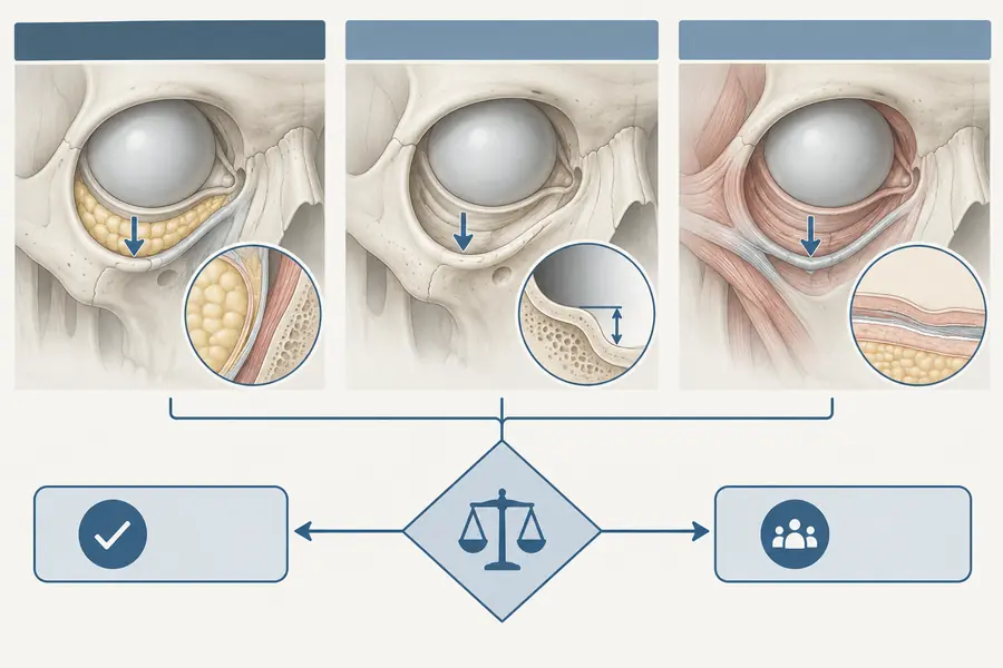
Anatomy of the temple and its danger zones
The layered anatomy of the temporal region, the vessels that make it hazardous, and why both blindness and venous embolism have been reported after treatment here.
The temple is often described as a straightforward region because the endpoint — restoring a hollow — looks simple. The anatomy underneath is not simple, and it is one of the few areas of the face where two different categories of serious complication have both been reported.
The layered arrangement
From surface to bone the temple is conventionally described in five layers: skin; subcutaneous fat; the superficial temporal fascia (also called the temporoparietal fascia, continuous with the SMAS below and with galea above); a loose areolar plane; and the deep temporal fascia, which splits over the upper part of the zygomatic arch into superficial and deep layers enclosing the superficial temporal fat pad. Beneath that lie temporalis muscle and periosteum.
Depth in the temple is therefore not a continuum. Each interface behaves differently, and the structures at risk change entirely depending on which one a needle tip has crossed. The two planes most commonly described for volume restoration are the supraperiosteal plane against bone and the subcutaneous plane immediately under skin. The layers between them contain the vessels.
The arterial consideration
The superficial temporal artery is the terminal branch of the external carotid. It runs within the superficial temporal fascia, dividing into frontal and parietal branches, and in many people it is palpable and visibly tortuous. Its course is variable enough that surface landmarks alone are unreliable.
The zygomatico-orbital artery arises from the superficial temporal artery and runs forward, superficially, above and roughly parallel to the zygomatic arch towards the lateral orbit. This is the branch that matters most, because it anastomoses with the periorbital circulation and therefore with branches of the ophthalmic artery — a vessel of the internal carotid system. That anastomotic connection is the anatomical basis for retrograde embolic travel from a temporal injection into the orbital circulation, and it places the temple on the published list of regions from which visual loss has been reported.
The venous consideration
The middle temporal vein runs within or immediately superficial to the deep temporal fascia, generally a short distance above the zygomatic arch, and it is a wide, thin-walled, low-pressure channel draining towards the internal jugular system. Filler entering it does not occlude a distal territory; it travels centrally. Case reports describe pulmonary embolism after temporal injection by this route. It is a mechanism unlike arterial occlusion in every respect — different presentation, different timing, and not something hyaluronidase into the face addresses.
Why this region collects reports
Most facial danger zones concentrate one failure mode. The temple concentrates two, in different layers, from a single entry point that offers little tactile feedback. The overlying tissue is thin, the vessels are variable, and the plane that feels “safe” by resistance alone may be exactly the plane containing the vein.
Imaging
Because the vascular course here is variable rather than merely deep, published anatomical work on the temporal region increasingly recommends ultrasound assessment before injection to identify vessel position in the individual patient rather than relying on averaged landmarks. The same literature notes that ultrasound is also the practical means of confirming which fascial plane a cannula or needle tip actually occupies. Neither point removes the risk; both change how it is estimated.