Injection anatomy review across the face
A revision note drawing the facial regions together: layers, fat compartments, retaining ligaments, the named danger zones and the carotid anastomosis behind them.
Facial anatomy is normally learned region by region. This note runs the other way, pulling the common structure out of the regional detail, because the principles that prevent complications are the same everywhere and only the named structures change.
Layers
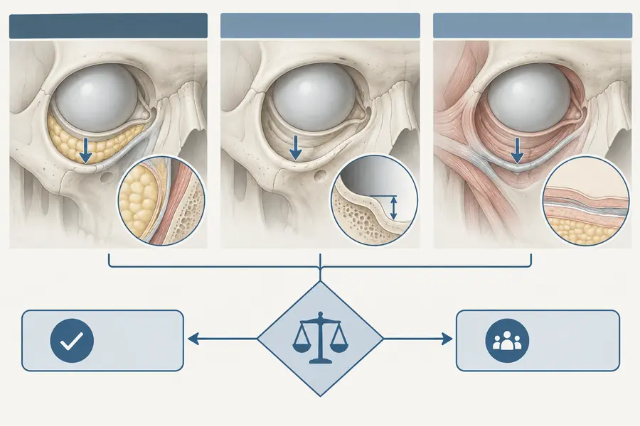
The face is arranged in five layers: skin; subcutaneous fat; the musculo-aponeurotic layer (SMAS, continuous with galea above and platysma below); a plane of loose areolar tissue and retaining ligaments; and the deep fascia and periosteum. The arrangement holds across the face, but the layers are not of equal thickness everywhere, and in some regions one of them is effectively absent — the eyelid has almost no subcutaneous layer at all.
Two consequences follow. First, depth is regional, not absolute: a stated distance from the skin means different things at the temple and at the chin. Second, most vessels of concern travel in predictable layers, which is what makes plane discipline a safety measure rather than a stylistic preference.
Compartments
Subcutaneous fat is not one continuous mass. It is divided into discrete compartments separated by septa, and these age at different rates — deep compartments deflating while superficial ones descend. This explains why volume placed at the site of a visible hollow often fails: the apparent defect is frequently the consequence of change in a neighbouring compartment.
Retaining ligaments
Osteocutaneous and fasciocutaneous ligaments tether the soft tissue envelope to the underlying skeleton — the zygomatic, mandibular, masseteric and tear trough attachments among them. They create the fixed points, and the visible grooves of the ageing face largely occur where tethered tissue meets untethered tissue that has descended.
For injection they matter twice. They define where product will and will not spread, and they mark the transitions where neurovascular structures characteristically cross between layers.
Danger zones
The published danger zones are not arbitrary. Each is a place where a named vessel is superficial, variable in course, or connected to the orbital circulation: the glabella, supratrochlear and supraorbital regions; the nasal dorsum and tip; the nasolabial fold and adjacent angular artery; the infraorbital and tear trough region; and the temple.
The literature on safe zones is the useful complement. Anatomical studies describing the sub-SMAS spaces identify planes that are relatively avascular and therefore comparatively tolerant, which is a more actionable formulation than a list of places to avoid.
The anastomosis
The single fact that unifies the danger zones: the face is supplied mainly by branches of the external carotid artery, while the eye is supplied by the ophthalmic artery from the internal carotid. The two systems anastomose freely around the orbit — through the angular, dorsal nasal, supratrochlear and supraorbital vessels, and laterally through zygomatico-orbital connections.
An injection made with enough pressure into a distal branch of the external carotid system can therefore drive material retrograde against flow, into the anastomosis, and forward into the ophthalmic circulation when pressure is released. That is the mechanism behind reported visual loss, and it is why glabellar and nasal injections carry ocular risk despite the distance involved.
The practical residue
Know which layer the needle is in. Know what runs in that layer here. Prefer low pressure and small increments, keep the tip moving, and treat every high-risk region as one where the individual anatomy may not match the diagram.