Neurosensory innervation
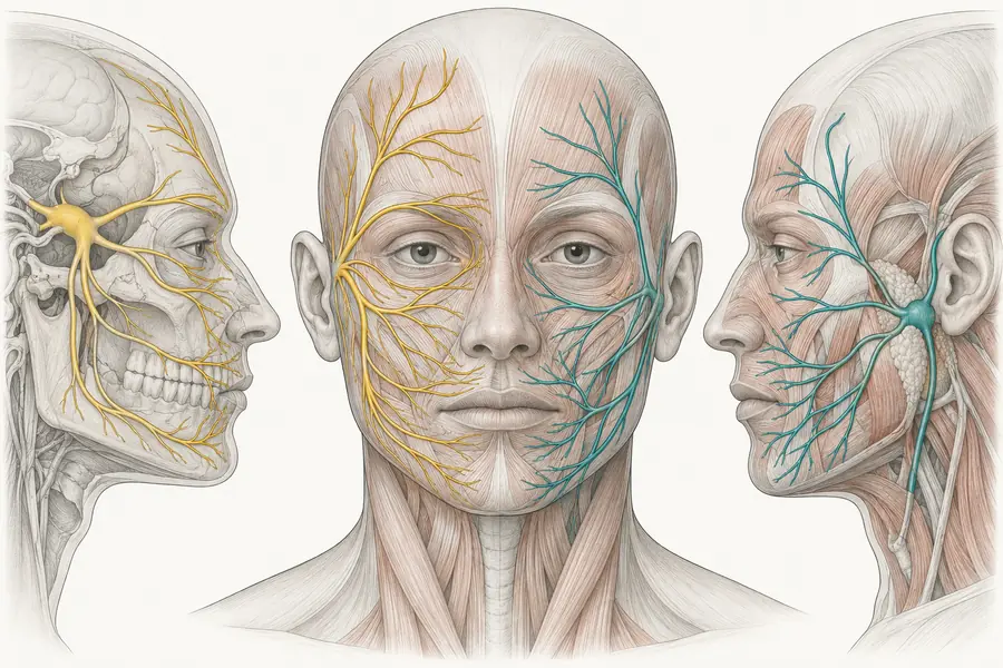
The sensory nerve supply of the face — chiefly trigeminal, with cervical plexus contribution — and the foramina where those nerves emerge and are vulnerable.
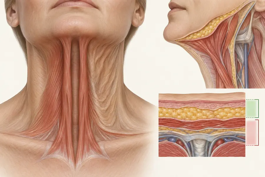
Sensation in the face comes almost entirely from the trigeminal nerve, with the skin over the mandibular angle and much of the neck supplied instead by the cervical plexus.
Three exit points matter practically because they are palpable, predictable and roughly aligned in the vertical mid-pupillary plane:
- the supraorbital foramen or notch at the superior orbital rim;
- the infraorbital foramen, about a centimetre below the inferior orbital rim;
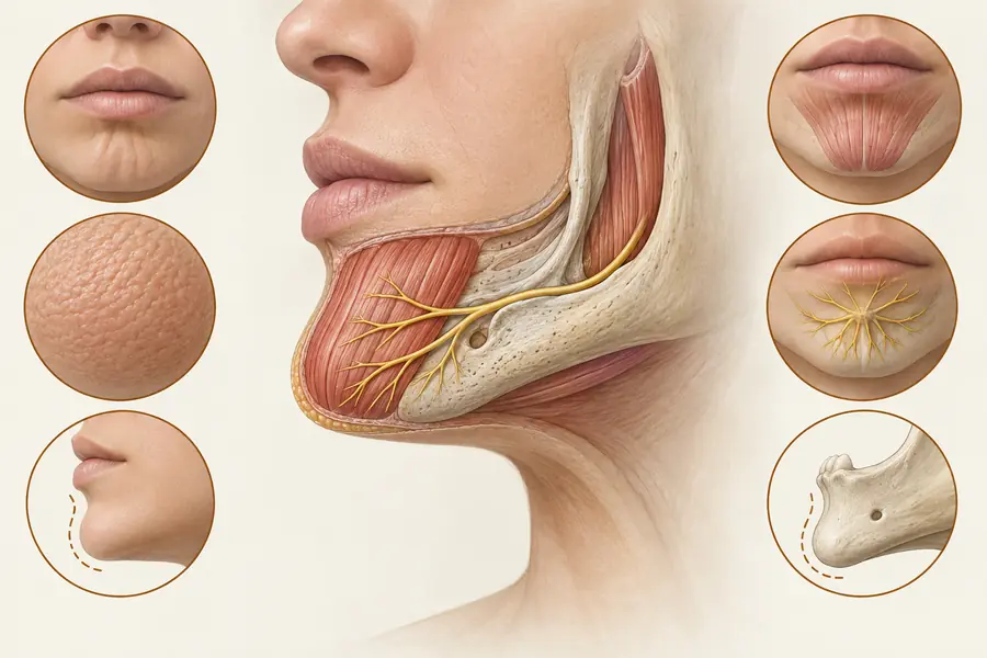
- the mental foramen, on the mandible between the premolars.
Each transmits a sensory nerve alongside an artery, and each is a site where injected volume can compress the nerve and produce numbness, paraesthesia or neuropathic pain.
There are two clinical uses for this map. It supports regional blocks: a small volume of local anaesthetic at these foramina anaesthetises a large territory, which is more comfortable than repeated infiltration. And it explains post-treatment sensory changes — numbness in a defined dermatome after infraorbital work points to compression at the foramen rather than to a generalised problem.
Sensory symptoms that persist beyond the expected resolution of swelling warrant review rather than reassurance.