External and internal carotid arteries
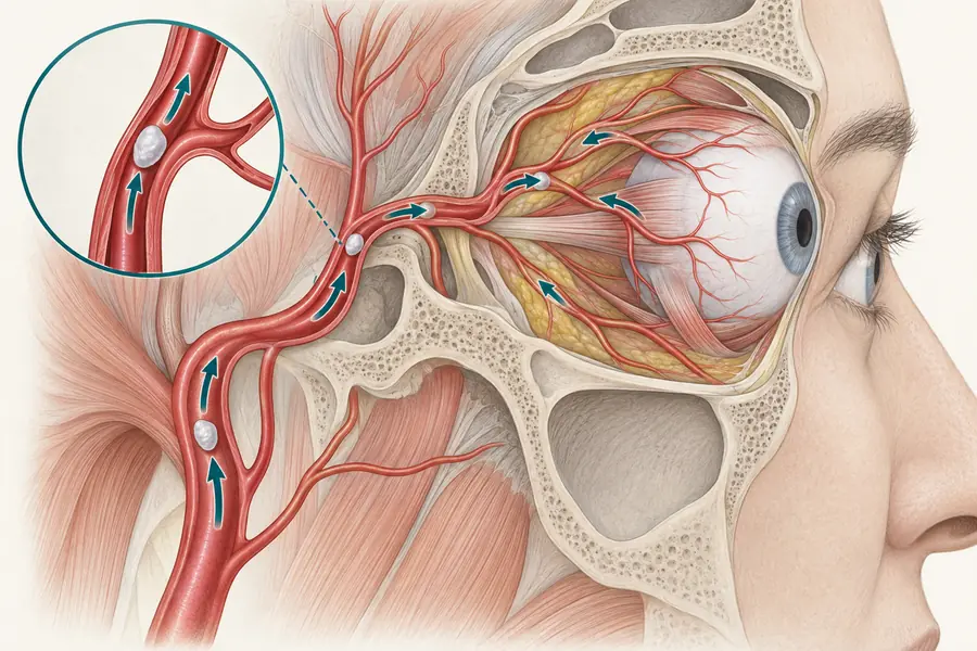
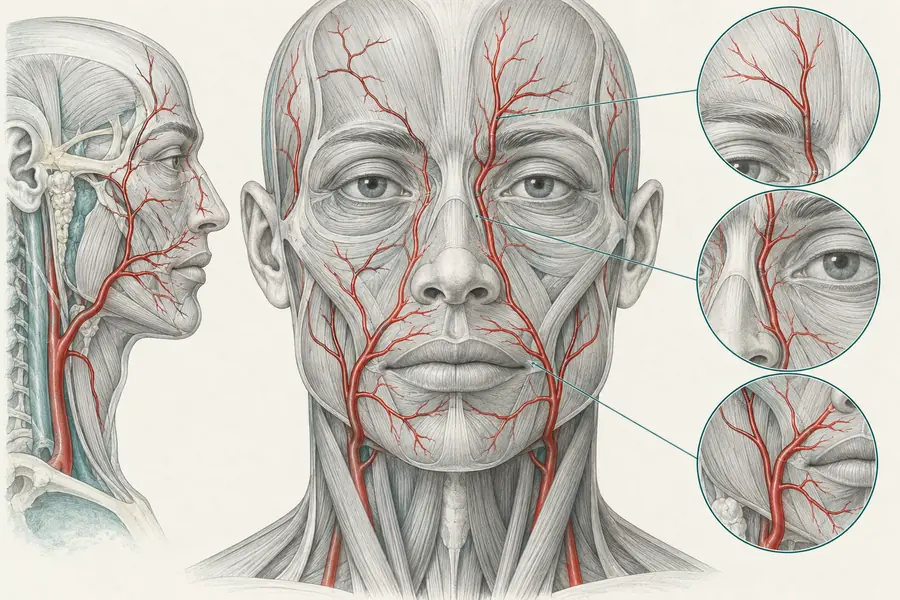
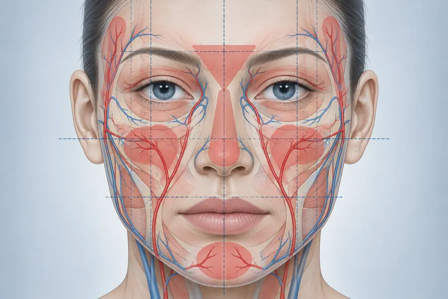
The two arterial systems supplying the head, which anastomose around the orbit — the anatomical basis of vision loss after facial filler injection.
The common carotid divides into an external and an internal carotid artery, and the relationship between the two territories is the single most important vascular fact in injectable practice.
The external carotid supplies the face through branches including the facial artery, which becomes the angular artery beside the nose, and the superficial temporal artery.
The internal carotid supplies the brain and, through the ophthalmic artery, the orbit and the eye. The ophthalmic artery’s terminal branches — supratrochlear and supraorbital — emerge onto the forehead at the orbital rim.
These systems are not separate. They anastomose, most importantly at the medial canthus where the angular artery meets the dorsal nasal branch of the ophthalmic. There are further connections across the nasal dorsum and in the temple.
The consequence is direct: material injected under pressure into an external carotid branch can travel retrograde past an anastomosis and, when normal flow resumes, be carried into the ophthalmic circulation. This is why glabella, nasal dorsum and periorbital regions account for the majority of reported blindness cases, and why no facial region can be described as vascularly isolated.