Tear trough assessment: the three causes of the hollow
The infraorbital hollow has three distinct origins that look alike at rest, and telling them apart determines whether filler is appropriate at all.
This address is preserved from the original site; the page covers the topic rather than any individual.
A hollow beneath the eye is a description of an appearance, not a diagnosis. At least three different anatomical situations produce something that looks similar in a static photograph, and they call for entirely different responses. The assessment, not the technique, decides the outcome.
Cause one: ligamentous tethering
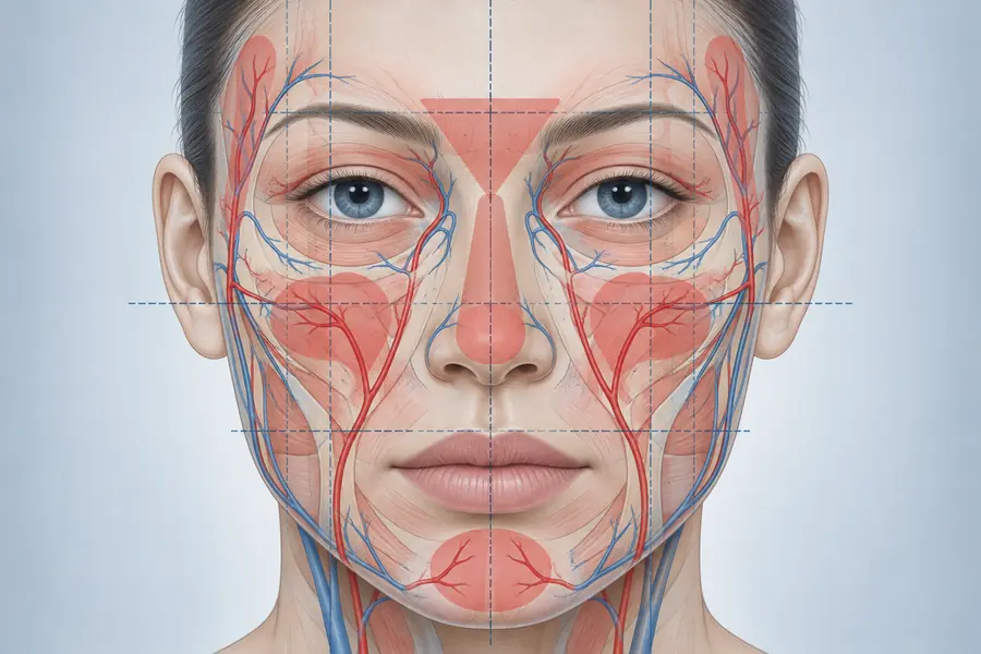
The tear trough proper corresponds to an osteocutaneous attachment running from the maxilla to the dermis along the inferomedial orbital rim. Where tissue is bound down, it cannot descend with the tissue around it, and a groove appears at the boundary.
This is a true tethered depression. It is present in youth in some people, it is visible in photographs from decades earlier, and its depth changes comparatively little with position or expression. Skin overlying it is thin, with minimal subcutaneous fat and orbicularis oculi immediately beneath — the least forgiving tissue on the face for anything placed superficially.
Cause two: midface deflation
Here the groove is a consequence of what has happened below it. Loss of volume in the medial cheek withdraws support from the infraorbital region, and the orbital rim becomes visible as a shadowed transition because the tissue that previously filled the contour beneath it has diminished.
The distinguishing feature is that the hollow is part of a continuous change over the upper midface rather than a discrete line. It generally coexists with flattening of the anterior cheek and a lengthening of the lid-cheek junction, and it is the cause most amenable to volume replacement — though usually to volume placed in the cheek rather than in the trough itself.
Cause three: pseudoherniated orbital fat
The third situation is not a hollow at all but a projection. Orbital fat pushes forward against a weakened orbital septum and produces a convexity above the rim; the groove below it is the boundary of that convexity, and it appears deeper because of what sits above it.
Telling them apart
Assessment is dynamic and positional, not photographic.
Change of posture is the most informative single manoeuvre. Fat pseudoherniation is gravity-dependent and reduces when the patient lies supine; a tethered groove does not change. Upward gaze and gentle globe pressure accentuate a herniated compartment.
Illumination from above exaggerates a convexity; light directed from below flattens the shadow of a true depression while a projection continues to cast one. Palpation distinguishes a firm, fixed, linear attachment from generalised softness of a deflated cheek. And a review of the patient’s earlier photographs indicates whether the feature is longstanding or recent.
Most patients present with a combination. The question is which element predominates.
Why the third is not a filler problem
Adding volume to an excess is an error of category rather than of technique. Placing product below a herniated fat compartment does not remove the convexity; it raises the surrounding contour so that the whole region projects, producing a heavier, fuller lower lid rather than a smoother one.
The tissue characteristics compound it. The region has thin skin and limited lymphatic drainage, so a hydrophilic gel placed there can generate sustained oedema that the patient reads as a poor result, and superficial placement risks a visible bluish discolouration. Pseudoherniation is a structural problem of the orbital septum, and its management belongs to a surgical conversation rather than an injectable one.
Recognising that at assessment is the whole of the skill. It is also why the honest answer at consultation is sometimes that the appearance in question is not something filler improves.